Back Of Skull Muscle Anatomy : Muscles Of The Head Learn Human Anatomy Neck Muscle Anatomy Human Anatomy Picture Muscle Anatomy : A collection of anatomy notes covering the key anatomy concepts that medical students need to learn.
Back Of Skull Muscle Anatomy : Muscles Of The Head Learn Human Anatomy Neck Muscle Anatomy Human Anatomy Picture Muscle Anatomy : A collection of anatomy notes covering the key anatomy concepts that medical students need to learn.. They don't move and united into a single unit. The splenius muscles originate at the midline and run laterally and superiorly to their insertions. Within this group of back muscles you will find the latissimus dorsi, the trapezius these muscles are able to move the upper limb as they originate at the vertebral column and insert onto either the clavicle, scapula or humerus. Posterior rami of the spinal nerves. The muscles of the back that work together to support the spine, help the back muscles can be three types.
The calf muscle, on the back of the lower leg, is actually made up of two muscles: For more in depth tutorials about the back muscles see my individual tutorials on the extrinsic back muscles and the intermediate and deep muscles. Muscles that act on the back. My name is alex, and i'm the owner and author of king of the gym. From an anatomical perspective, the skull is divided into two parts:
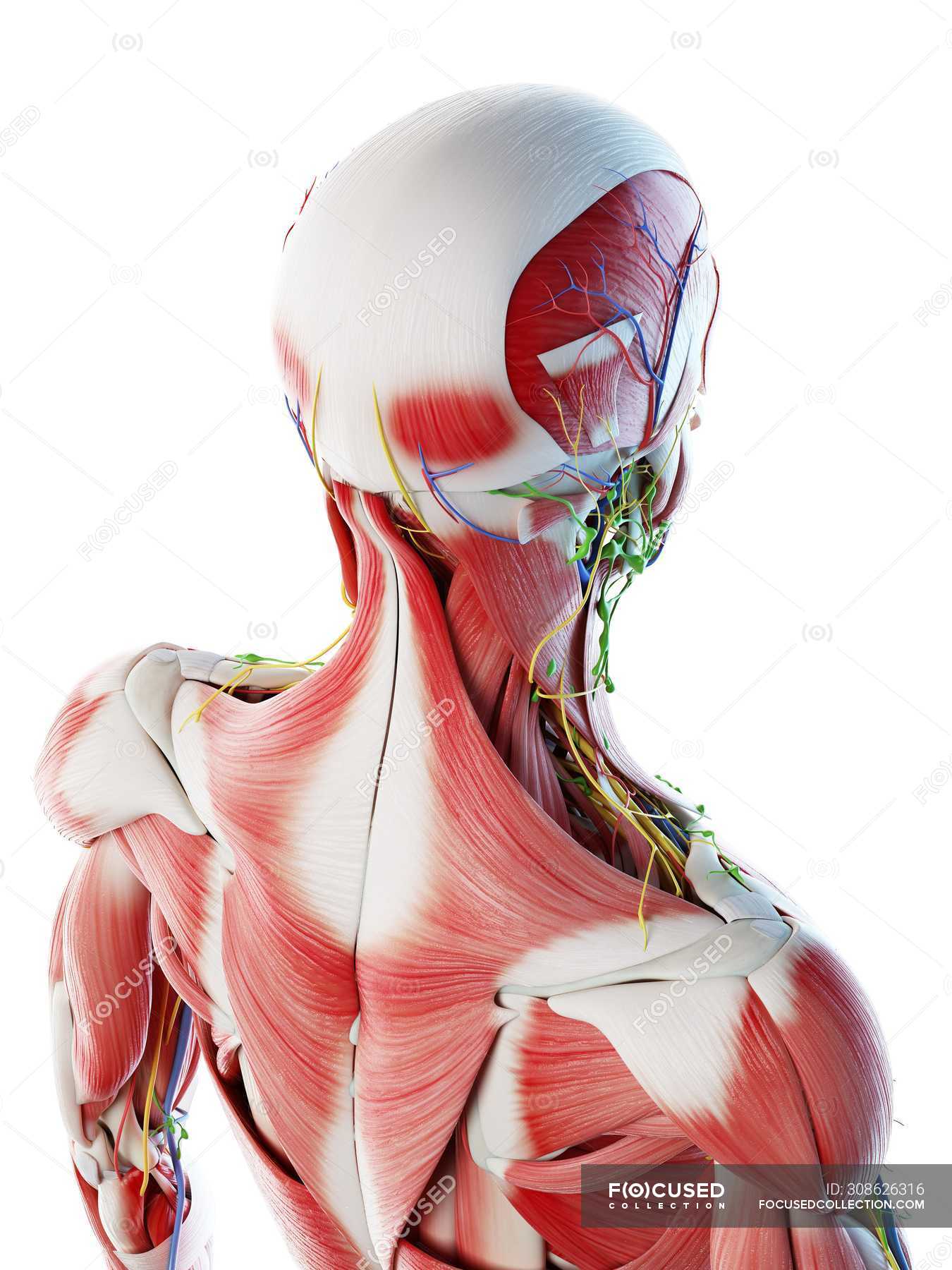
Male Back Neck And Head Muscles Computer Illustration Anatomical 3d Model Stock Photo 308626316 from st.focusedcollection.com I think we have a respectable sense of how muscles contract on the molecular level let's take a step back now and just understand how muscles look at least structurally or how they relate to things that we normally associate with muscles so let me draw. Some expressions are done by one or two muscles, while others take several muscles at once. Musculoskeletal, cardiovascular, nervous, respiratory, digestive, urogenital (male and female), endocrine, lymphatic, eye and ear. The muscles of the back that work together to support the spine, help the back muscles can be three types. Join our newsletter and receive our free ebook: Through a simple and intuitive interface it is possible to observe systems: They are the muscle group of the back responsible for extension, adduction, and rotation of the upper limbs. The muscles of the thoracic area lie deep to the functional anatomy:
For more in depth tutorials about the back muscles see my individual tutorials on the extrinsic back muscles and the intermediate and deep muscles.
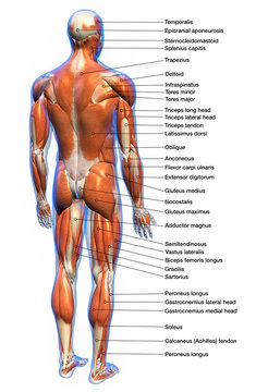
Some expressions are done by one or two muscles, while others take several muscles at once. Join our newsletter and receive our free ebook: From an anatomical perspective, the skull is divided into two parts: The muscles of the thoracic area lie deep to the functional anatomy: This is a table of skeletal muscles of the human anatomy. We study anatomy at the practical anatomy class we study the human body. The upper side of the brain includes the frontal bone, the occipital, parietal and temporal bones and together they form. Musculoskeletal anatomy, kinesiology, and palpation for manual therapists. Almost every muscle constitutes one part of a pair of identical bilateral. For more in depth tutorials about the back muscles see my individual tutorials on the extrinsic back muscles and the intermediate and deep muscles. Occipital bone of the skull, ligamentum nuchae, and the spinou… spine & acromion of the scapula, and lateral 1/3 of the clavic… Front view of muscles, skeleton, organs, nervous system. The gastrocnemius is the larger calf muscle, forming the bulge visible calf muscle rupture:
The muscles of the skull and face are divided into two groups. The upper back is a complex area containing a number of muscles that perform various actions on the scapulae shoulder blades and humerus. Learn about anatomy back muscles with free interactive flashcards. The gastrocnemius is the larger calf muscle, forming the bulge visible calf muscle rupture: Guide to mastering the study of anatomy.
17 085 Best Back Muscles Anatomy Images Stock Photos Vectors Adobe Stock from t3.ftcdn.net A collection of anatomy notes covering the key anatomy concepts that medical students need to learn. The upper back is a complex area containing a number of muscles that perform various actions on the scapulae shoulder blades and humerus. The muscles of the skull and face are divided into two groups. Guide to mastering the study of anatomy. The skull or known as the cranium in the medical world is a bone structure of the head. The splenius muscles originate at the midline and run laterally and superiorly to their insertions. The physicians originally studying human anatomy thought the skull looked like an apple. The galea joins the frontalis muscle belly anteriorly to the occipitalis muscle belly posteriorly.
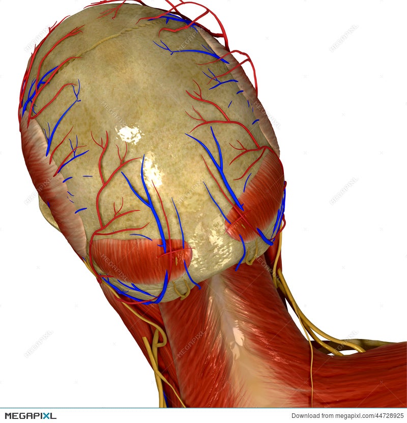
At the lower end is the nuchal ridge where some neck muscles attach.
The cranium and the mandible. They move the head in every direction, pulling the skull and jaw towards the shoulders, spine, and scapula. The gastrocnemius is the larger calf muscle, forming the bulge visible calf muscle rupture: I started this website back in late 2009 during college, and it has been my. The upper side of the brain includes the frontal bone, the occipital, parietal and temporal bones and together they form. A collection of anatomy notes covering the key anatomy concepts that medical students need to learn. Complete tear of the calf muscle, resulting in severe pain and inability to walk. The muscles of the skull and face are divided into two groups. The simplest way to make the difference between the head and the back of the head or occipital bone has four aesthetic bony regions. Guide to mastering the study of anatomy. The skull performs vital functions. The upper back is a complex area containing a number of muscles that perform various actions on the scapulae shoulder blades and humerus. The calf muscle, on the back of the lower leg, is actually made up of two muscles:
The occipital bone of the skull. The cranium and the mandible. The upper back is a complex area containing a number of muscles that perform various actions on the scapulae shoulder blades and humerus. The thick muscles of the heart contract to pump blood out and then relax to let blood back in after it has circulated through the body. Skull reshaping is done on any of the structures that lie above the face.
Skull With Muscles And Nerves Back View Illustration 44728925 Megapixl from images.megapixl.com Front view of muscles, skeleton, organs, nervous system. The muscles of the skull and face are divided into two groups. The upper back is a complex area containing a number of muscles that perform various actions on the scapulae shoulder blades and humerus. Discover the muscle anatomy of every muscle group in the human body. Some common muscles involved with neck pain include the sternocleidomastoid, trapezius, levator scapulae the trapezius is a large surface muscle that spans from the base of the skull down the spine to the mid back, as well as out. This is why raising your eyebrows can create the appearance that the back of the head is moving. We study anatomy at the practical anatomy class we study the human body. It supports and protects the face and the brain.
The occipital bone of the skull.
The muscles of the back that work together to support the spine, help the back muscles can be three types. A skull consists of the frontal, temporal, parietal and occipital bones. The muscles of mastication are responsible for the movement of the mandible during mastication (chewing). My name is alex, and i'm the owner and author of king of the gym. The back muscle anatomy is made up of large and small muscle groups all working harmony to help with those everyday movements. The superficial back muscles are the muscles found just under the skin. The gastrocnemius is the larger calf muscle, forming the bulge visible calf muscle rupture: For more in depth tutorials about the back muscles see my individual tutorials on the extrinsic back muscles and the intermediate and deep muscles. They don't move and united into a single unit. A collection of anatomy notes covering the key anatomy concepts that medical students need to learn. Musculoskeletal, cardiovascular, nervous, respiratory, digestive, urogenital (male and female), endocrine, lymphatic, eye and ear. Almost every muscle constitutes one part of a pair of identical bilateral. Occipital bone of the skull, ligamentum nuchae, and the spinou… spine & acromion of the scapula, and lateral 1/3 of the clavic…


0 Komentar